 Official websites use .gov
A .gov website belongs to an official government organization in the United States. Official websites use .gov
A .gov website belongs to an official government organization in the United States. |
 Secure .gov websites use HTTPS
A lock or https:// means you’ve safely connected to the .gov website. Share sensitive information only on official, secure websites. Secure .gov websites use HTTPS
A lock or https:// means you’ve safely connected to the .gov website. Share sensitive information only on official, secure websites. |
Solutions today for reefs tomorrow
Dark spot disease (DSD) is a non-lethal coral disease that is characterized by lesions of purple or brown discoloration. Dark spot disease is known to affect 21 scleractinian coral species and is most prevalent on the reef-building species Siderastrea siderea. Since it was first reported in Colombia in the 1990s, DSD has been observed throughout the wider Caribbean, Brazil, and in the Indo-Pacific.


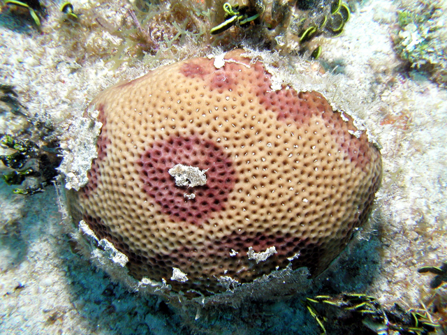
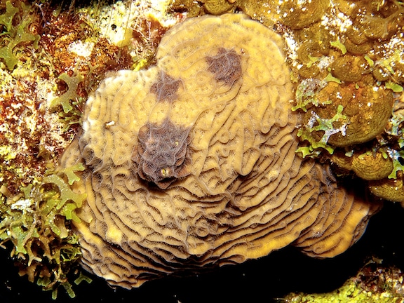
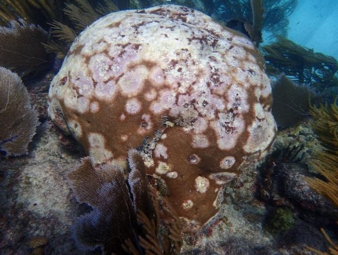
Dark spot disease lesions appear as multifocal to coalescing areas of purple or brown discoloration, ranging in size from 1-5cm, that can be located diffusely over the entire colony. The discolored areas can be round, oblong, or irregular spots with smooth or undulating margins.
Dark spot disease lesions can progress over time, though the progression rate typically remains low at <2cm/ month. In some cases, the center of lesions can exhibit subacute tissue loss with algal overgrowth (indicating the characteristically slow lesion progression rate) or can have a depressed relief. It is currently unknown whether this feature is caused by slower growth or active decalcification in affected areas.
Dark spot disease lesions are not lethal and can persist on a colony for years, with a notable exception being a mortality event of Agaricia agaracites in Colombia in 2004.


The precise cause of DSD is not currently known. Some microbiological studies have found that DSD-affected tissues host a different bacterial consortium than that found on healthy tissues, including a notably high abundance of the bacterium Vibrio carchariae, but inoculating a healthy coral with the bacteria isolated on a diseased coral does not produce a DSD lesion. This, coupled with the ineffective treatment with antibiotics, suggests that a bacterial pathogen is unlikely to be the cause of DSD.
Histopathological studies on DSD from the Indo-pacific and Florida have identified communities of fungus within the skeletons underlying DSD lesions. The fungal mats, known as “endolithic fungi,” because of their residence within the skeleton of a coral, are dense within the skeleton and appear to affect the architecture of adjacent coral tissues. This, coupled with the characteristically slow and chronic lesion behavior, suggest that a fungal pathogen may be the cause of DSD, though this needs to be confirmed with culturing and inoculation studies.
The overall impact of DSD on reef coral cover and biodiversity is low. Dark spot disease lesions are non-lethal, and while they can be chronic and persist for multiple years, there is also some evidence of healing and tissue regeneration.
Borger, J. L. (2003). Three scleractinian coral diseases in Dominica, West Indies: Distribution, infection patterns and contribution to coral tissue mortality. Revista de Biologia Tropical, 51(SUPPL. 4).
Borger, J. L. (2005). Scleractinian coral diseases in south Florida: Incidence, species susceptibility, and mortality. Diseases of Aquatic Organisms, 67(3). https://doi.org/10.3354/dao067249
Borger, J. L., & Steiner, S. C. C. (2005). The spatial and temporal dynamics of coral diseases in Dominica, West Indies. Bulletin of Marine Science, 77(1).
Brandt, M. E., & Mcmanus, J. W. (2009). Disease incidence is related to bleaching extent in reef-building corals. Ecology, 90(10). https://doi.org/10.1890/08-0445.1
Cervino, J., Goreau, T. J., Nagelkerken, I., Smith, G. W., & Hayes, R. (2001). Yellow band and dark spot syndromes in Caribbean corals: Distribution, rate of spread, cytology, and effects on abundance and division rate of zooxanthellae. Hydrobiologia, 460. https://doi.org/10.1023/A:1013166617140
Correa, A. M. S., Brandt, M. E., Smith, T. B., Thornhill, D. J., & Baker, A. C. (2009). Symbiodinium associations with diseased and healthy scleractinian corals. Coral Reefs, 28(2). https://doi.org/10.1007/s00338-008-0464-6
Cróquer, A., & Weil, E. (2009). Changes in Caribbean coral disease prevalence after the 2005 bleaching event. Diseases of Aquatic Organisms, 87(1–2). https://doi.org/10.3354/dao02164
Francini-Filho, R. B., Moura, R. L., Thompson, F. L., Reis, R. M., Kaufman, L., Kikuchi, R. K. P., & Leão, Z. M. A. N. (2008). Diseases leading to accelerated decline of reef corals in the largest South Atlantic reef complex (Abrolhos Bank, eastern Brazil). Marine Pollution Bulletin, 56(5). https://doi.org/10.1016/j.marpolbul.2008.02.013
Garzón-Ferreira, J., Gil-Agudelo, D. L., Barrios, L. M., & Zea, S. (2001). Stony coral diseases observed in southwestern Caribbean reefs. Hydrobiologia, 460. https://doi.org/10.1023/A:1013133818360
Gil-Agudelo, D. L., & Garzón-Ferreira, J. (2001). Spatial and seasonal variation of dark spots disease in coral communities of the Santa Marta area (Colombian Caribbean). Bulletin of Marine Science, 69(2).
Gil-Agudelo, D. L., Smith, G. W., Garzón-Ferreira, J., Weil, E., & Petersen, D. (2004). Dark Spots Disease and Yellow Band Disease, Two Poorly Known Coral Diseases with High Incidence in Caribbean Reefs. In Coral Health and Disease. https://doi.org/10.1007/978-3-662-06414-6_19
Gochfeld, D. J., Olson, J. B., & Slattery, M. (2006). Colony versus population variation in susceptibility and resistance to dark spot syndrome in the Caribbean coral Siderastrea siderea. Diseases of Aquatic Organisms, 69(1). https://doi.org/10.3354/dao069053
Kaczmarsky, L. T., Draud, M., & Williams, E. H. (2005). Is there a relationship between proximity to sewage effluent and the prevalence of coral disease? Caribbean Journal of Science, 41(1).
Voss, J. D., & Richardson, L. L. (2006). Coral diseases near Lee Stocking Island, Bahamas: Patterns and potential drivers. Diseases of Aquatic Organisms, 69(1). https://doi.org/10.3354/dao069033
Ward, J. R., Rypien, K. L., Bruno, J. F., Harvell, C. D., Jordán-Dahlgren, E., Mullen, K. M., Rodríguez-Martínez, R. E., Sánchez, J., & Smith, G. (2006). Coral diversity and disease in Mexico. Diseases of Aquatic Organisms, 69(1). https://doi.org/10.3354/dao069023
Weil, E., and Croquer, A. (2009) Spatial variability in distribution and prevalence of Caribbean scleracintion coral and octocoral diseases. I. Community-level analysis. Diseases of Aquatic Organisms 83, 195-208.
Weil, E., Smith, G., & Gil-Agudelo, D. L. (2006). Status and progress in coral reef disease research. In Diseases of Aquatic Organisms (Vol. 69, Issue 1). https://doi.org/10.3354/dao069001
Work, T. M., Aeby, G. S., Stanton, F. G., & Fenner, D. (2008). Overgrowth of fungi (endolithic hypermycosis) associated with multifocal to diffuse distinct amorphous dark discoloration of corals in the Indo-Pacific. Coral Reefs, 27(3). https://doi.org/10.1007/s00338-008-0374-7